
Advanced Office-Based Stress Echocardiography Services in Fort Wayne, Angola, Peru & Lagrange, IN
Exercise treadmill test (Stress Test)
This test uses a treadmill machine which progressively increases in speed and elevation while the patient is attach to an EKG machine. The patient is exercised in order to get an assessment of his exercise capacity and also to evaluate for blockage of the heart arteries (coronary artery disease), which is detected by the EKG changes while he is running on the treadmill. The test also checks for the capacity of the heart to increase its rate as more workload is put on it (chronotropic response). The test also is able check for exercise-induced arrhythmias.
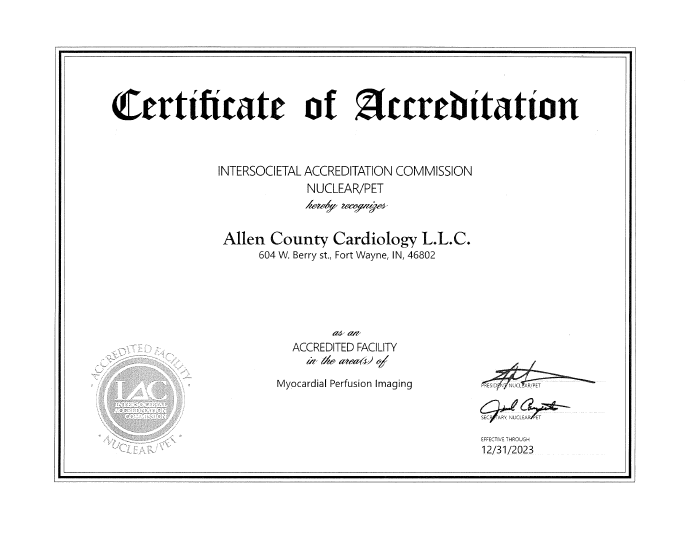
Nuclear stress testing (Exercise Myoview or Pharmacological Myoview Imaging)
This test involves injection of a radiopharmaceutical agent (usually Myoview or Cardiolite) during a treadmill or under a pharmacological agent (Lexiscan). Subsequently, the comparison of images of the heart between the stress and rest can unmask coronary artery obstruction and also reveal previous heart muscle damage (myocardial infarction). This technique also helps the physician to assess the global and regional functions of the heart (left ventricular ejection fraction). Undoubtedly, this carries a better sensitivity and specificity as compared to the exercise treadmill test and usually is the test recommended for high risk patients and also for patients who are post-stent and post-bypass surgery (coronary artery bypass graft).

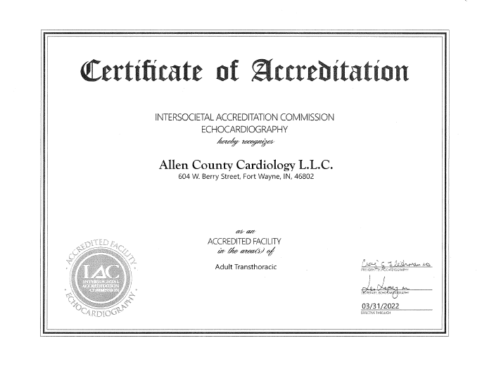
Transthoracic echocardiography with Doppler
This is an imaging study in which ultrasound waves are used to create two-dimensional pictures of the cardiac muscle, cardiac valves, and other structures such as pericardium and great vessels of the heart. Doppler evaluation helps to further detect valvular defects and other defects of the heart. Fluid around the heart can also be detected by this study.
Stress echocardiography
Stress echo combines echo during peak treadmill and comparing it with rest echo. This test can unmask coronary artery disease due to obstructed plaque. If done properly, this test also carries a higher sensitivity and specificity as compared to plain exercise treadmill tests; however, major limitations include the quality of the study, which may be impaired in some patients who have a big body size and chronic lung disease.
Holter monitoring
This is a test used to diagnose cardiac arrhythmias which may be accounting for the patient’s symptoms, such as fainting or palpitations. The patient carries a Holter box connected to electrodes on the chest usually for 24 hours, and sometimes up to 48 hours. This is a noninvasive test.
Event recorders
Similar to a Holter monitor, event recorders usually involve wearing the recording machine for a longer duration of up to 30 days. It is usually used to monitor heart rhythm disorders that occur in a sporadic fashion.
Carotid Doppler
This is an imaging technique which uses ultrasound waves to detect blockages and the degree of thickness of the intima of the carotid arteries (arteries which supply the brain and are present in the neck).
Lower/upper extremity arterial Doppler and venous Doppler
In this test, arteries and veins of the upper and lower extremities are visualized for obstruction and clots by ultrasound waves.
Lipid Testing
Fingerstick test used to check cholesterol levels.
Abdominal Aortic Aneurysm Detection
Ultrasound waves are used to detect the size of the abdominal aorta. The test is usually recommended for male smokers 65-years or older or for patients with a family history of clinical symptoms that need this test.
Renal Duplex Scan
Ultrasound waves are used to detect any significant narrowing of renal arteries. The test is usually recommended for patients in whom blood pressure is difficult to control and also if there is a clinical suspicion of renal artery narrowing.
INR Testing
Finger stick blood is used to check adequacy of blood thinning by Coumadin (warfarin).
Advanced Cardiac Diagnostics at Allen County Cardiology
At Allen County Cardiology, we understand how critical early detection is when it comes to heart health. That’s why our cardiologist in Fort Wayne, IN, offers advanced diagnostic services like EKG testing and heart scans to help pinpoint heart issues before they become serious. These noninvasive procedures provide vital insights into your cardiovascular condition and are key to creating an effective treatment plan tailored to you.
EKG Testing
EKG testing is one of the most effective ways to monitor your heart’s rhythm and electrical activity. Whether you're experiencing symptoms like chest pain, dizziness, or palpitations, or simply taking a proactive approach to your heart health, our cardiologist in Fort Wayne, IN, uses EKGs to detect irregularities such as arrhythmias, past heart attacks, and more. At Allen County Cardiology, we make EKG testing a comfortable, quick, and essential step in understanding your heart’s performance.


Heart Scan
A heart scan offers a deeper look into potential coronary artery disease by detecting calcium deposits in the arteries. This test is especially valuable for patients with risk factors like high blood pressure, high cholesterol, or a family history of heart disease. Our expert cardiologist in Fort Wayne, IN, uses this high-tech imaging tool to uncover early warning signs of heart trouble—even before symptoms appear. When it comes to your heart, a heart scan could make all the difference in catching issues early. Don’t wait to take control of your heart health. Trust the experienced team at Allen County Cardiology. Call us today to schedule your appointment and consult with a leading cardiologist in Fort Wayne, IN, who puts your heart first.
Heart disease in Fort Wayne, IN continues to be a leading cause of illness and complications for both men and women. At Allen County Cardiology, our board-certified cardiologists provide hospital-based procedures and individualized treatment plans for patients at all stages of cardiovascular disease (CVD). Whether you are newly diagnosed or managing ongoing cardiovascular issues, we’re here to help you take control of your heart health today.
Call us now at
(260) 489-4100 to schedule an appointment.
